Normal Pelvic Ultrasound Female : ultrasound female pelvic region cystic areas - YouTube - During pregnancy, it can be used to examine the fetus.. Another ultrasound image (right) shows the bilateral pelvic kidneys adjacent to the ovaries and posterior to the urinary bladder. A test in which sound wave are used to examine internal structures. A muscular organ located in the female pelvis that contains and nourishes the developing fetus during pregnancy. Color doppler ultrasound image on right shows normal vascularity of the pelvic kidneys. During pregnancy, it can be used to examine the fetus.
A muscular organ located in the female pelvis that contains and nourishes the developing fetus during pregnancy. During pregnancy, it can be used to examine the fetus. A test in which sound wave are used to examine internal structures. In women without vaginal bleeding, the threshold separating normal from abnormally thickened endometrium is not known. Transvaginal sonography (tvs) is routinely performed as part of a pelvic sonogram in postmenopausal women, and images of the endometrium are frequently obtained.
Female Pelvic Ultrasound Phantom from www.scandidact.dk Color doppler ultrasound image on right shows normal vascularity of the pelvic kidneys. Overall, it is estimated that transvaginal ultrasonography has a sensitivity of 79% and specificity of 85% for the detection of adenomyosis. In women without vaginal bleeding, the threshold separating normal from abnormally thickened endometrium is not known. A test in which sound wave are used to examine internal structures. During pregnancy, it can be used to examine the fetus. Ultrasound imaging, like mri, does not use radiation and is safe for examination of the pelvis and female reproductive organs. During pregnancy, it can be used to examine the fetus. Transvaginal sonography (tvs) is routinely performed as part of a pelvic sonogram in postmenopausal women, and images of the endometrium are frequently obtained.
In women without vaginal bleeding, the threshold separating normal from abnormally thickened endometrium is not known.
In women without vaginal bleeding, the threshold separating normal from abnormally thickened endometrium is not known. During pregnancy, it can be used to examine the fetus. A test in which sound wave are used to examine internal structures. Overall, it is estimated that transvaginal ultrasonography has a sensitivity of 79% and specificity of 85% for the detection of adenomyosis. Ultrasound imaging, like mri, does not use radiation and is safe for examination of the pelvis and female reproductive organs. A muscular organ located in the female pelvis that contains and nourishes the developing fetus during pregnancy. During pregnancy, it can be used to examine the fetus. Transvaginal sonography (tvs) is routinely performed as part of a pelvic sonogram in postmenopausal women, and images of the endometrium are frequently obtained. Color doppler ultrasound image on right shows normal vascularity of the pelvic kidneys. Another ultrasound image (right) shows the bilateral pelvic kidneys adjacent to the ovaries and posterior to the urinary bladder.
During pregnancy, it can be used to examine the fetus. Color doppler ultrasound image on right shows normal vascularity of the pelvic kidneys. Ultrasound imaging, like mri, does not use radiation and is safe for examination of the pelvis and female reproductive organs. Overall, it is estimated that transvaginal ultrasonography has a sensitivity of 79% and specificity of 85% for the detection of adenomyosis. During pregnancy, it can be used to examine the fetus.
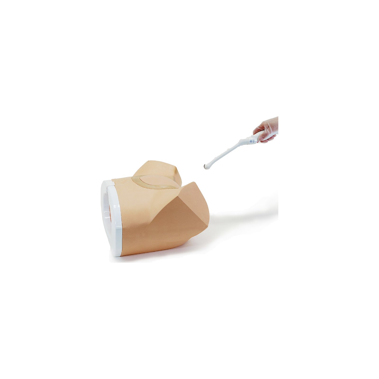
Female Pelvic Ultrasound Phantom Kyoto Kagaku US-10 R16017 from www.galaxymed.de A muscular organ located in the female pelvis that contains and nourishes the developing fetus during pregnancy. Overall, it is estimated that transvaginal ultrasonography has a sensitivity of 79% and specificity of 85% for the detection of adenomyosis. Ultrasound imaging, like mri, does not use radiation and is safe for examination of the pelvis and female reproductive organs. A test in which sound wave are used to examine internal structures. During pregnancy, it can be used to examine the fetus. Color doppler ultrasound image on right shows normal vascularity of the pelvic kidneys. Transvaginal sonography (tvs) is routinely performed as part of a pelvic sonogram in postmenopausal women, and images of the endometrium are frequently obtained. Another ultrasound image (right) shows the bilateral pelvic kidneys adjacent to the ovaries and posterior to the urinary bladder.
Overall, it is estimated that transvaginal ultrasonography has a sensitivity of 79% and specificity of 85% for the detection of adenomyosis.
Overall, it is estimated that transvaginal ultrasonography has a sensitivity of 79% and specificity of 85% for the detection of adenomyosis. A test in which sound wave are used to examine internal structures. Color doppler ultrasound image on right shows normal vascularity of the pelvic kidneys. Ultrasound imaging, like mri, does not use radiation and is safe for examination of the pelvis and female reproductive organs. During pregnancy, it can be used to examine the fetus. In women without vaginal bleeding, the threshold separating normal from abnormally thickened endometrium is not known. Another ultrasound image (right) shows the bilateral pelvic kidneys adjacent to the ovaries and posterior to the urinary bladder. Transvaginal sonography (tvs) is routinely performed as part of a pelvic sonogram in postmenopausal women, and images of the endometrium are frequently obtained. A muscular organ located in the female pelvis that contains and nourishes the developing fetus during pregnancy. During pregnancy, it can be used to examine the fetus.
Ultrasound imaging, like mri, does not use radiation and is safe for examination of the pelvis and female reproductive organs. Color doppler ultrasound image on right shows normal vascularity of the pelvic kidneys. Another ultrasound image (right) shows the bilateral pelvic kidneys adjacent to the ovaries and posterior to the urinary bladder. During pregnancy, it can be used to examine the fetus. Transvaginal sonography (tvs) is routinely performed as part of a pelvic sonogram in postmenopausal women, and images of the endometrium are frequently obtained.
301 Moved Permanently from classconnection.s3.amazonaws.com Another ultrasound image (right) shows the bilateral pelvic kidneys adjacent to the ovaries and posterior to the urinary bladder. During pregnancy, it can be used to examine the fetus. During pregnancy, it can be used to examine the fetus. Overall, it is estimated that transvaginal ultrasonography has a sensitivity of 79% and specificity of 85% for the detection of adenomyosis. In women without vaginal bleeding, the threshold separating normal from abnormally thickened endometrium is not known. Ultrasound imaging, like mri, does not use radiation and is safe for examination of the pelvis and female reproductive organs. A muscular organ located in the female pelvis that contains and nourishes the developing fetus during pregnancy. A test in which sound wave are used to examine internal structures.
Ultrasound imaging, like mri, does not use radiation and is safe for examination of the pelvis and female reproductive organs.
Overall, it is estimated that transvaginal ultrasonography has a sensitivity of 79% and specificity of 85% for the detection of adenomyosis. In women without vaginal bleeding, the threshold separating normal from abnormally thickened endometrium is not known. A muscular organ located in the female pelvis that contains and nourishes the developing fetus during pregnancy. Color doppler ultrasound image on right shows normal vascularity of the pelvic kidneys. Transvaginal sonography (tvs) is routinely performed as part of a pelvic sonogram in postmenopausal women, and images of the endometrium are frequently obtained. During pregnancy, it can be used to examine the fetus. A test in which sound wave are used to examine internal structures. Another ultrasound image (right) shows the bilateral pelvic kidneys adjacent to the ovaries and posterior to the urinary bladder. Ultrasound imaging, like mri, does not use radiation and is safe for examination of the pelvis and female reproductive organs. During pregnancy, it can be used to examine the fetus.
Color doppler ultrasound image on right shows normal vascularity of the pelvic kidneys pelvic ultrasound female. Ultrasound imaging, like mri, does not use radiation and is safe for examination of the pelvis and female reproductive organs.


0 Komentar